Blog

  • Intoxication: working out possible ingested dose

    Intoxication: working out possible ingested dose

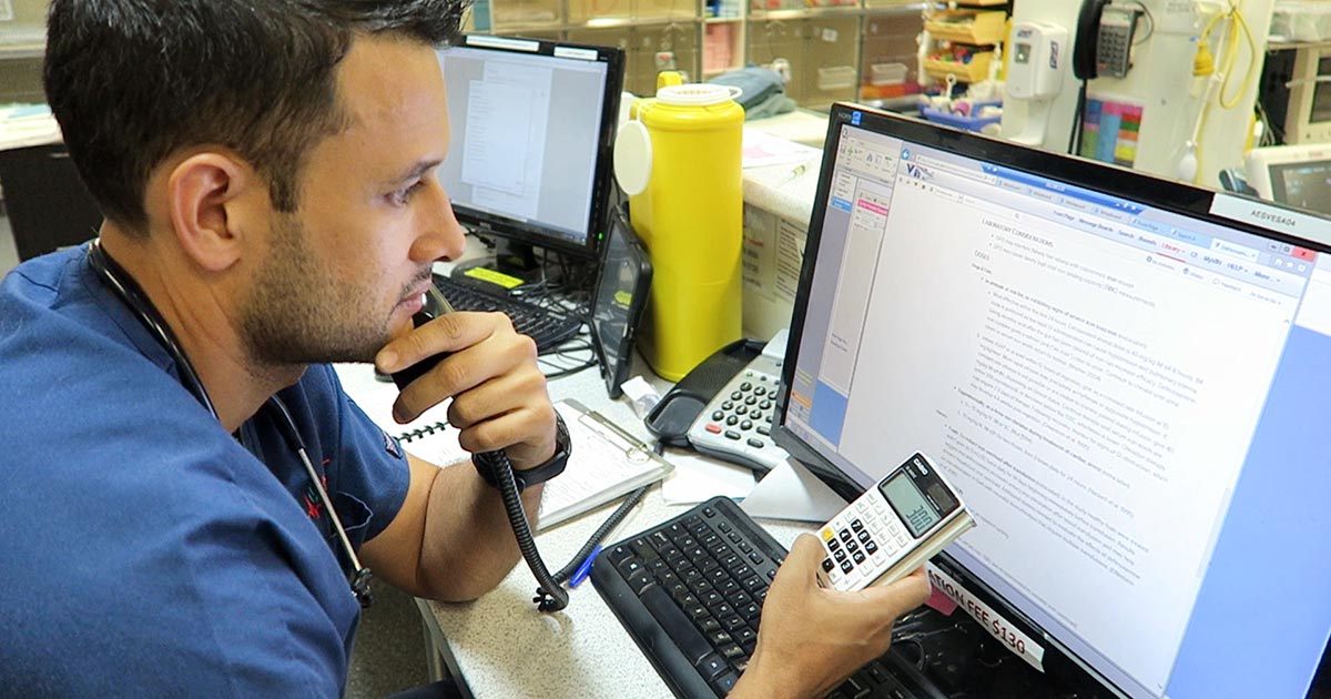
    We frequently field telephone calls from owners concerned about their pet being intoxicated or having access to a toxic compound.

    These are the list of questions I always ask owners:

    What is your pet doing?

    The main reason I ask this question first is to determine if the pet’s life is in danger. If the pet is seizing, collapsed, neurological, bleeding or having difficulty breathing then they need to come into practice immediately.

    What led to the suspicion of toxic exposure?

    This can help provide useful background information.

    What is the product?

    In some situations, owners can tell you accurately over the telephone what they think they have been exposed to.

    Asking them to bring the packaging, and whatever is remaining of the toxin, with them can help determine a possible dose they have been exposed to.

    When did this occur?

    A timeline, and when they think the pet could have been exposed to the toxin, is critical as it can help put presenting clinical signs into perspective.

    I always ask if they could have had prior exposure to the toxin. An example where this may be important is with rodenticides.

    What have you done in response to this?

    Owners may have tried to address the situation themselves, using information gained from the internet.

    Attempts to induce emesis can also make pets incredibly ill and result in neurological signs.

    Have they passed faeces or vomitus with the toxin?

    If the answer is yes, ask them to bring the pet into the practice. This can help identify the toxin; some baits are coloured and can easily be seen. These samples may even be able to be sent away for further testing.

    Do you have any other pets that may have also had access to the toxin?

    Other pets that may have had access will need to be seen in practice as well. A classic example is a multi-dog household where one pet is the scavenger. Owners may neglect to inform you their other pets may have been the culprits, but did not because they assumed it was the one with the history of being a scavenger.

    Next week, we will cover the decontamination steps owners can carry out at home.

  • Icteric serum

    Icteric serum

    The final discolouration of the serum we are going to cover is icteric serum.

    Icteric serum
    Icteric serum is caused by the presence of excess bilirubin in the blood stream.

    Icteric serum is caused by the presence of excess bilirubin in the blood stream as a result of increased production (pre-hepatic) or inappropriate excretion (hepatic and post-hepatic).

    The most common cause of pre-hepatic icterus is haemolytic anaemia, while hepatic disease and biliary tract obstruction are the most common causes for hepatic and post-hepatic icterus, respectively.

    Tips on where to start

    If icterus and concurrent anaemia exist, my first suspicion would be some kind of pre-hepatic cause. The most common causes are immune-mediated haemolytic anaemia and infectious haemolytic anaemia, such as haemotropic mycoplasma and babesiosis.

    Other causes can include snake envenomation and oxidative injury from heavy metal toxicity or onion ingestion.

    Regarding hepatic and post-hepatic causes, unfortunately it is not always clear-cut. Both are commonly associated with elevation in both alanine transaminase (ALT) and alkaline phosphatase (ALKP), and, although no specific changes are pathognomonic for hepatic or post-hepatic disease, the pattern of change may help identify the origin of the cause. ALT is released from the inside of hepatocytes, and in higher amounts when cell damage occurs.

    Hepatic hints

    Some pointers on what you can do to help differentiate:

    • Compare the ALT and ALKP elevation; if one is in order of magnitudes higher than the other then it can help point to an origin.
    • If the cause is of hepatic origin, one would expect the ALT to be significantly more elevated than the ALPK. Likewise, this is usually true in reverse for post-hepatic causes. However, it should be noted in chronic hepatic diseases, where active damage to hepatocytes is comparatively lower, a mild increase in ALT and marked increase in ALPK does not preclude disease of hepatic origin. Therefore, biopsies should always be used for definitive diagnosis.
    • If other biochemistry parameters such as albumin, glucose and cholesterol are low, or prolonged clotting times are present, the case for a hepatic origin is strengthened.
    • The gallbladder and bile duct can be assessed using abdominal ultrasonography. The presence of a dilated bile duct, or evidence supportive of pancreatitis, is highly suggestive of a post-hepatic cause.

    Finally, it is important to be aware of the impact on hyperbilirubinaemia on laboratory testing. Hyperbilirubinaemia generally causes decreased cholesterol, triglyceride, creatinine, lipase, total protein and gamma-glutamyltransferase levels.

  • Lipaemia – the bane of biochemistry

    Lipaemia – the bane of biochemistry

    Last week we covered haemolysed samples – this week we’re looking at lipaemic samples.

    Lipaemic samples are caused by an excess of lipoproteins in the blood, creating a milky/turbid appearance that interferes with multiple biochemical tests and can even cause haemolysis of red blood cells.

    lipaemic sample
    A severely lipaemic sample (red arrow). IMAGE: eClinPath.com (CC BY-NC-SA 4.0).

    Lipaemia can follow recent ingestion of a meal – especially one high in fat. Although not pathognomonic for any diseases, its presence can help increase the suspicion of certain diseases, including:

    • pancreatitis
    • diabetes mellitus
    • hypothyroidism
    • hyperadrenocorticism
    • primary hyperlipidaemia (in some specific breeds, such as the miniature schnauzer)

    It warrants further investigation in patients that have been ill and inappetent.

    Irksome interpretations

    Lipaemia can dramatically impact laboratory testing and is often troublesome in critically ill patients, making interpretation of biochemistry particularly difficult, if not impossible.

    Lipaemia can affect different analysers in different ways, most commonly causing:

    • Falsely increased calcium, phosphorus, bilirubin, glucose and total protein (via refractometer) and some liver parameters such as alkaline phosphatase, alanine aminotransferase, aspartate aminotransferase, haemoglobin concentration, and mean corpuscular haemoglobin concentration.
    • Falsely decreased sodium, potassium, chloride, albumin and bicarbonate.

    Tube tips

    Assessment of a centrifuged haematocrit tube before running a biochemistry panel can help reduce wasted biochemistry consumables.

    If the sample is lipaemic in the haematocrit tube then maybe try some of the following tips.

    • If blood tests are planned in advance, try fasting the patient beforehand for 12 to 24 hours.
    • Repeat sampling a couple of hours later may yield a less lipaemic sample.
    • Collecting and centrifuging a larger amount of blood (3ml to 5ml, for example) can sometimes yield enough clear sample between the lipid layer and red blood cells.
    • Refrigeration of the sample can help the separation.
    • Extract lipids using polar solvents, such as polyethylene glycol.
    • Centrifugation at higher than normal speeds (if possible) can also assist in clearing the layer.
  • PCV/total solids interpretation: serum colour

    PCV/total solids interpretation: serum colour

    When interpreting the often misinterpreted and underused PCV and total solids test, it is important to take note of the serum colour as this may give clues into the diagnosis.

    PCV tubes
    Normal serum colour (left) compared to a patient with immune-mediated haemolytic anaemia. The serum is haemolysed and anaemia is present.

    The most common abnormalities seen in clinic are icteric, haemolysed and lipaemic serum.

    Clear serum can also be of importance – especially when you interpret it with blood counts and urine colour.

    Haemolysis

    The most common abnormality of serum colour changes is haemolysis. In my experience, the most common cause is suboptimal collection technique. To confirm this, simply collect another sample and repeat.

    If it is repeatable, and concurrent anaemia or pigmenturia is present, it warrants further investigation.

    Intravascular haemolysis can be caused by:

    • immune-mediated haemolytic anaemia
    • blood transfusion reactions
    • infectious diseases such as Mycoplasma haemofelis, Babesia canis, Ehrlichia canis, FeLV and others
    • Heinz bodies from the ingestion of heavy metal, onions or paracetamol
    • hypophosphataemia
    • macroangiopathic disease (neoplasia, for example)
    • envenomation – typically, snake bites

    Testing issues

    Haemolysis can also affect other laboratory testing. It can lead to an artefactual increase in glucose, phosphorus, bilirubin, total protein, fructosamine and triglycerides, and a decrease in sodium (pseudohyponatraemia), cholesterol, calcium, potassium and albumin.

    Extravascular haemolysis often does not cause haemolysed serum as it is generally slower and the body is able to clear the haemoglobin before it can lead to discolouration of the serum.

  • ‘Long COVID’ within the veterinary industry

    ‘Long COVID’ within the veterinary industry

    Despite that all COVID-19 restrictions are due to be removed as early as the end of the month, the long-term impacts of this pandemic have yet to ease and will likely be sending ripples through many professions for the foreseeable future.

    The virus has already left lasting changes to the landscape of the veterinary industry – not only in how it operates, but in how new professionals are taught from the ground up. Long gone are the days of packed-out waiting rooms and lecture theatres.

    The more things change

    Even with restrictions out of the way, it’s thought that measures put in place over the past two years to reduce viral transmission in our practices may remain the “new normal”, with clients asked to wait outside until necessary, and often with only a single owner asked to come in with their pet at any one time.

    Several vet schools now operate under the banner of “blended learning”, whereby the curriculum is taught in a mixture of face-to-face content, live and online lectures, alongside pre-recorded, virtually accessible resources.

    In the face of recurrent industrial action – coupled with the rising, desperate demand for new veterinary professionals – online teaching may offer a convenient, long-term solution.

    Fallen behind

    As with all change, there are pros and cons, although one area I think is yet to be fully addressed is the issue of EMS backlogs.

    There are current backlogs in the NHS, the postal system and in many other major industries throughout the country, and I don’t believe the veterinary student training system has escaped unaffected.

    Car park consults may remain the “new normal” for some practices. Image © Eva / Adobe Stock

    Exaggerating social inequity

    For a good 12 to 18 months, many practices – especially small independents – were forced to shut their doors to both pre-clinical and clinical placements – and although EMS requirements were lowered for all year groups due to graduate by 2023, every succeeding year must complete the formerly required 26 weeks.

    This has led to an overwhelmingly large number of students competing for a progressively smaller number of placement opportunities.

    This makes it particularly difficult for students who lack their own transport, or the funds to travel far from their university or hometown, meaning there may be growing social inequity in the variety and quality of experience vet students are able to obtain.

    Better the devil you know

    Not only this, but large veterinary conglomerates may have the infrastructure to accommodate larger numbers of students, and new graduates may want to “stick with what they know” and, therefore, be less likely to consider signing contracts with smaller, independent practices.

    There is also the argument that, with the rise of online teaching and decrease in student-lecturer contact, work experience has never been such a valuable tool in supplementing a student’s learning.

    With the demand for placements higher than ever before, and veterinary practices sometimes two or three times busier than before the pandemic, the financial cost of EMS for some students has never been greater.

    Essential experiences

    Confidence, satisfaction and a balanced education behind all new graduates serves the interest of both individual professionals, and the profession as a whole.

    In my opinion, there has never been a greater need for a review of the need for higher education funding for veterinary students, to allow every student access to a large variety of work experience and the opportunity to see what different avenues are available to them after graduation.

  • PCV/total solids: getting the most from simple test

    PCV/total solids: getting the most from simple test

    The PCV and total solids (TS) test is simple, yet informative – but is often misinterpreted or underused.

    Table 1. Changes that can be found on a PCV/TS and possible causes (click to view).
    Table 1. Changes that can be found on a PCV/TS and possible causes (click to zoom).

    It is important to remember all test results need to be interpreted in light of the patient’s history, presenting clinical signs and general physical examination findings.

    The various changes that can be found on a PCV/TS, and the possible causes, are detailed in Table 1. Many of the differentials can be included or excluded based on the history, clinical signs and examination findings.

    Misconceptions

    I would like to highlight some common misconceptions I find with PCV/TS interpretation.

    A normal PCV/TS means the patient cannot be dehydrated

    The concept all dehydrated patients will have an elevated PCV/TS is inaccurate. Patients will have to be severely dehydrated to see an elevation in both PCV/TS.

    Dehydration should be based primarily on physical examination findings, not based primarily on PCV/TS results.

    A patient with a normal PCV could not have lost blood as the PCV should be low

    Patients can have acute whole blood loss, which is not reflected in the PCV at presentation. This could have been caused by a number of reasons – for example:

    • the extravascular fluid has not yet shifted down the hydrostatic pressure gradients
    • the patient has not ingested water since the time of blood loss
    • IV fluid has not been given to correct the hypovolaemia

    Once the fluid shifts, the patient drinks water or IV fluids are administered, the PCV/TS will drop due to haemodilution.

    An elevated PCV means the patient is dehydrated

    PCV tubes
    PCV tubes.

    This is probably the most common change I see in my patients – and it is not because they are all dehydrated.

    The most common cause of this change is stress-induced splenic contraction. The spleen stores red blood cells. Under the influence of adrenalin, the smooth muscle in the spleen contracts and the stored red blood cells are pushed into circulation.

    The next most common cause would be haemorrhagic gastroenteritis, where a fluid shift into the gastrointestinal tract has occurred.

    Assessment of the serum colour can also provide valuable information. White or lipaemic serum can cause artifactually high TS. Haemolysed serum with a low PCV can indicate a haemolytic anaemia.

    Next time you perform a PCV/TS, look at Table 1 and consider the other differentials for your results.

  • Euthanasia (part 2): caring for the patient

    Euthanasia (part 2): caring for the patient

    Last month we discussed the importance of caring for clients during the process of euthanising their much-loved pet. This month, we focus on your patient.

    The goals of euthanasia are always to make it as painless, fearless and stress-free as possible for the patient.

    Pain relief

    Most patients presented for euthanasia are either suffering from chronic, terminal or traumatic disease.

    The first thing I like to do is ensure the patient’s pain is managed. This usually means providing opioid pain relief. Methadone is my opioid of choice. Butorphanol provides minimal pain relief, but is excellent for mild sedation.

    Next, if your patient is in shock, you need to try to alleviate some of it through IV fluid resuscitation. This is important as poor circulation will slow the process when you administer the euthanasia solution.

    Calm and stress-free

    One of the most important goals in the euthanasia process is to have the patient as calm and stress-free as possible.

    If the patient is stressed or anxious, some sedation may be required. Diazepam or acepromazine are good choices, depending on the condition of the patient, of course, and, together with the opioid you have administered already for pain relief, will help calm the patient. Try to avoid using medetomidine as a sedative in all but the most fractious of patients, as it causes peripheral vasoconstriction that will make IV catheter placement difficult.

    The aim of sedation is to relax your patient as much as possible without rendering them unresponsive to owners when it is time to say goodbye. This can be tricky as every patient responds differently to sedation, so you must make a point of warning owners the sedation may make their pets very sleepy.

    IV catheter

    Where possible, I avoid performing euthanasia without first placing an IV catheter. It makes the delivery of the euthanasia so much smoother. Including an extension to the administration line also allows you to stand a little away from the patient and their owners to give them a little privacy while you administer the euthanasia solution.

    Once your patient is sedated and an IV catheter placed, I set up comfortable bedding in the room where I will perform the euthanasia and bring the patient to the room. The amount of euthanasia solution I have with me always exceeds how much I think I will need; the last thing I want is to have to leave the owner and patient to get more euthanasia solution.

    Once I have administered the euthanasia solution, I check the heart and once that stops completely, I then tell the owners their pet’s heart has stopped, which they understand as their pet has passed.

    Performing euthanasias is the one of the hardest parts of our job, but also a privilege we hold as vets, so I hope my tips will help make the process easier for you, your client and, most importantly, your patient.

  • Prophylactic mental health

    Prophylactic mental health

    Many facets of mental health are comparable to physical illness/fitness, and I’ve gained a lot of introspection on how to keep myself mentally healthy and happy on the vet course from lessons I’ve learned from my physical training.

    Since joining the gym a couple months before the pandemic hit (as futile as that turned out to be) to focus on my physical health, I’ve noticed a profound increase in my mental health and my general mood.

    There’s a lot that links mental and physical health that we’re barely scratching the surface of. Endorphins released while exercising improve your mood and reduce anxiety, wearing out your body a little more during the day helps you sleep better at night, and building a routine can certainly be beneficial in a course as time-management crucial as veterinary medicine.

    Getting physical

    Having said that, I think there’s a lot more to it when it comes to prophylactic mental health.

    When I started weightlifting, for example, it was easy to feel like more was better – to the point where I was putting in more time in the gym, but seeing less results.

    As counterintuitive as this sounds, your body needs time to rest and recover before starting back fresh with renewed levels of energy. Usain Bolt isn’t trying to break records every single day, and being at 110% all the time sounds exhausting… and is probably also impossible.

    The same can be said for working and revising. If you have an inbound deadline or exam, your impulse is likely to think that the more you work, the better your grade. But trust me, that’s just not always the case – and I learned this the hard way.

    Heed the warning signs

    After suffering from burnout in my first year – after three months straight of revision (which I began four months before exams) – I had to accept that, long term, high intensity just isn’t sustainable.

    Recently, I’ve been able to apply the warning signs I’ve learned to pick up on at the gym to my work on the vet course.

    If I’m noticing that I’m not motivated to work out, that the weight I lifted easily last week now feels super heavy, or my muscles are sore for longer, I know it’s time to tone it down. Similarly, if I can’t bring myself to stare at another textbook, if a fact I previously knew now keeps escaping me, or if I’m noticing longer periods of bad moods and shorter periods of feeling happy, I know that something needs to change.

    For the former problem, I take a couple days off, do some stretching, and give myself time to heal. For the latter, the process is much the same, rest, relaxation and doing my best to switch my mind off – whether that’s TV, a good book, or, ironically, a good gym session.

    Preventive approach

    With the busy work life that comes with being a vet, I think the luxury of being able to go to the gym three or four times a week falls by the wayside, despite even the best of intentions.

    I know of a lot of professionals who’ve given up hobbies like running or hiking because there simply aren’t the hours in the day.

    However, giving our medical staff the time to take care of their physical health could go a long way to lessening the mental health crisis that has existed in the veterinary community for decades.

  • Euthanasia (part 1): caring for the client

    Euthanasia (part 1): caring for the client

    Euthanasia is a big part of our work as veterinarians. Working in an emergency setting, it is something I have to face on every shift.

    It doesn’t get any easier no matter how many times I have to do it, but I have fine-tuned my approach over the years so each euthanasia process runs as smoothly as possible, with minimal additional stress to both patient and client.

    This month, I will talk about taking care of your client.

    Communication is key

    dog and owner
    Euthanasia is a big part of our work as veterinarians.

    The most important aspect of taking care of your client in this difficult time is to make sure you really focus on communicating clearly, effectively and, most importantly, with sincere empathy.

    First, I listen to their concerns, and why they have made the difficult decision to euthanise their pet.

    Quality of life decisions can be a very grey area, and sometimes what you think may be manageable as a veterinarian can be a huge quality of life concern for a pet owner.

    A prime example is osteoarthritis in older dogs. You may assess them as being clinically well except for some difficulty walking, but the client sees their pet every day and notices the struggles they go through.

    Euthanasia is a difficult conclusion for them to come to and, in most instances, I will defer to the client when it comes to assessing the quality of life of their pet.

    Quality assessment

    One way I help clients assess their pet’s quality of life is by asking them about a few aspects of it, including:

    • Can your pet do the things that make them happy?
    • Do they spend more days sad, depressed and ill compared to the number of days they are bright, happy and eating?
    • Is your pet in pain? Is this pain manageable?

    Confirmation

    Once a client has expressed they want to euthanise their pet, I always try to confirm three things:

    1. That they have actually decided to euthanise their pet. I frame the question like: “So, my understanding from our conversation is that you have made the decision to euthanise Fluffy today?” Sometimes, when you ask this question, the client reveals they have not actually come to that decision yet, which means you will need to backtrack a little and guide them through the decision process again.
    2. Whether they would like to be present for the euthanasia.
    3. How they would like us to handle the after care.

    I also always try to manage all documentation and finances before the euthanasia so the clients will be in a position to leave immediately after the procedure, meaning they can begin to grieve rather than have to do paperwork. The only exception to this is when the patient is in a critical condition, meaning euthanasia cannot wait.

    Explaining the process

    Try not to perform the euthanasia in your consult room or in the main treatment areas – if you have a private room for euthanasias, that is the most ideal. This is important especially if the client comes back in the future with another pet or a new pet. They often find it difficult to walk into your consult room and be reminded of the euthanasia of their beloved pet.

    I like to give clients some time to spend alone with their pet to say their goodbyes in private. When I come back into the room, I start by explaining the process of the euthanasia, covering the following things every single time:

    • Euthanasia is an overdose of an anaesthetic agent
    • dog collarThe process is quick – 10 to 20 seconds
    • It is completely painless
    • The pet doesn’t close their eyes afterwards
    • The pet can have a couple deep breaths and muscle tremors
    • The pet can release their bowels and bladder (especially important to warn of this if the clients want to hold their pet)
    • Lastly, if their pet came into the hospital in shock and obtunded, where I have fluid resuscitated them, meaning they are now more bright and alert, I warn the clients that despite their pet looking better, the underlying disease remains the same.

    Once the euthanasia is performed, I again ask if the client wants to spend a little more time in private with their pet. Finally, when the client leaves, they typically will say “thank you”.

    Whatever you do, do not say something like “my pleasure” or “you’re welcome” like you would for a vaccination consult – this is a natural response, but would be a terrible faux pas. I simply say “I’m very sorry for your loss. Take care for now and let us know if we can help in any way”.

    Next month, I will talk about taking care of your patient throughout the euthanasia process.

  • Fluid therapy part 4: ongoing losses

    Fluid therapy part 4: ongoing losses

    This month, we will look at the final part of a fluid therapy plan – accounting for ongoing losses. This can be challenging, but some general rules can be helpful.

    Regular assessment is essential to track patients' response.
    Regular assessment is essential to track patients’ responses.

    First, let’s recap the four parts of a fluid therapy plan:

    1. Perfusion deficit
    2. Hydration deficit
    3. Maintenance requirements
    4. Ongoing losses

    When considering ongoing losses, try to not forget about patients with pre-existing polyuric diseases; chronic renal failure is a prime example. Patients with dehydrated chronic renal failure are unlikely to suddenly regain concentrating ability. Polyuria should be considered as an ongoing loss.

    Other conditions that may result in additional urinary fluid losses include post-obstructive diuresis, diabetes mellitus, hyperadrenocorticism and hyperthyroidsim.

    How much to add?

    This is the tricky part. I often add an additional half to one maintenance and frequently reassess clinical parameters, or if a urinary catheter is placed matching ins and outs.

    Gastrointestinal tract losses can be collected and weighed; 1g of vomitus or diarrhoea can be roughly equivalent to 1ml of water.

    Fluid removed from drains placed in cavities or wounds should also be measured and accounted for.

    Remember the key point is regular assessment of the patient’s hydration status, from repeat clinical exams, to track their response. Don’t forget regular retesting of electrolytes – for example, every 12 to 24 hours for patients on IV fluids and not eating.