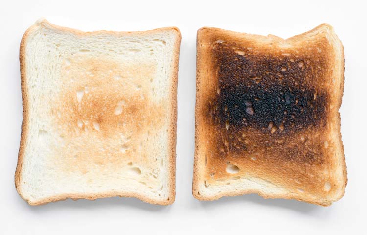
If you find rules of exposure confusing, then a simple tip is to think of it like toast…
Nurses are usually great at radiographic technique, with the flip side that vets are often poor… if you find rules of exposure confusing, then a simple tip is to think of it like toast: overcooked toast burns and goes black.
If your KV is set too high for the area being imaged, the film will be over exposed (dark) and demonstrate a flat, grey background. That is, of course, if you have not pushed it to far so as to black out (burn out) the image. If the KV is too low, the dense areas being imaged will not be penetrated. The film, or parts of it, will be white.
If the MAS is too high, the film will be over exposed (dark) demonstrating a black background. If the MAS is too low, the film will be white and washed out.
Lastly, if you change one factor (MAS or KV) you may need to change the other so as to balance the film density.
So:
If your films are dark and grey reduce the KV.
If your films are dark and black reduce the MAS.
If your films are light with no bony outlines clearly seen, increase the KV.
If your films are light but the bony outline is there, increase the MAS.
A couple of weeks ago, I received the phone call I’ve been dreading since moving away to university.
My horse had had an accident in the field, hurt her leg, and the vet was on the way. That’s all the information I received until the next call, with the vet on the other end.
“Communication within the tarsal joint… leg swinging… don’t think the long bone is actually fractured but significant damage to tendons at the back… rapid respiratory rate.”
As soon as I knew which way the conversation was going, I barely heard the rest.
My girl, who I’d had for 10 years. My girl, who’d been passed from pillar to post before we gave her the stable long-term home she’d never had. My girl, who had taught me to ride by being an infuriatingly awkward cow at the best of times.
My girl, who, when in the mood, was unbeatable and with whom I achieved a national title. My girl, who was the only one I trusted not to hurt me after my four-week stint in hospital when another horse landed on me. My 21-year-old girl, who’d been steadily getting stiffer from arthritis over the last few months. My girl, who, when I last rode about a week before this incident, was 10 times better than she’d been in a long time.
“My girl, who, when in the mood, was unbeatable and with whom I achieved a national title.”
My girl was about to be shot…
All the vet language stopped making sense, the clinician rationale went out the window. I just needed to know one thing, vet to vet student: is this fair? Could it wait five hours for me to tear down the M6 to say a final goodbye or would even that be an unnecessary amount of suffering?
I think I already knew the answer.
I felt utterly helpless and beyond reason for the following days, but as the shock wore off I was able to consider things retrospectively.
Having spoken to the family that were with her at the time, I’ve gathered a bit more information and been able to convince myself it was the right and only decision.
RIP Pepsi.
I’ve seen many animals have to be euthanised, for varying reasons. Some cases were more upsetting than others, but, for the most part, I’ve been able to detach myself from it – always telling myself it was for the best, in the animals’ interest for welfare reasons, and that there were no alternatives.
They say clients will only take in a small proportion of bad news. Now I know what that means. All the vet talk just went straight over my head, and the only thing I really got was that there was only one way it was going. This has outlined the importance of clear and concise communication when delivering bad news to my own clients in the future.
In communication skills classes, we’re told to encourage owners to bring someone with them who can write down key points and ensure they understand before proceeding. Now I appreciate the value of this so much more, having been the receiver instead of the bearer of bad news.
While the pain is still raw, I think I can take something from this to help me be more empathetic and ensure I communicate effectively in the future.
You can steal all the ham sandwiches you want now, Pepsi.
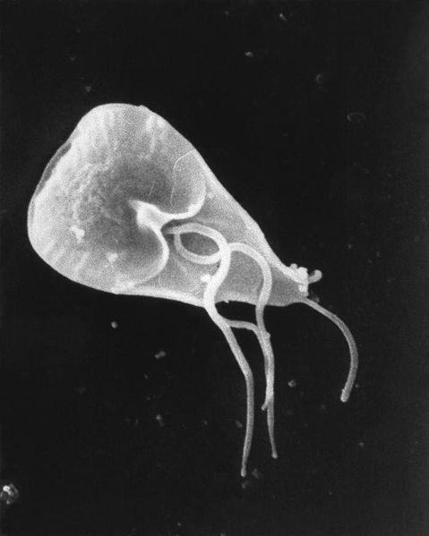
A flagellated Giardia lamblia protozoan parasite viewed with a scanning electron micrograph (SEM).
Giardia lamblia is a protozoan parasite found in the small intestine of vertebrates. The most common cause of transmission in cats is faecal-oral, but cats can also become infected by drinking water containing the infective cysts.
Most cats are asymptomatic, although they may keep passing on cysts for months or years. Clinical signs are most likely to be seen in younger animals from multi-cat households or environments.
If large numbers of trophozoites develop the cat will develop symptoms, which include:
Faecal flotation in zinc sulfate solution may be used to detect cysts. Three stool samples should be studied over a period of 7-10 days before a definite diagnosis is made. Alternatively, ELISA (enzyme-linked immunosorbent assay) can be used.
Giardiasis (also known as beaver fever) can be treated with metronidazole, furazolidone or fenbendazole.
Angiostrongylus vasorum is the most common cause of haemorrhagic cerebrovascular accidents in dogs.
Bleeding in the brain or spinal cord can cause neurological symptoms. Craniotentorial bleeding can cause epileptic seizures, paresis and abnormal postural reactions.
In cases of cerebellar bleeding, hypermetria, vestibular symptoms and opisthotonus are observed, and if the brainstem is affected, abnormalities of the cranial nerves can be seen.
Inflammation, hypoxia and parasitic emboli may cause neurological signs too.
It is worth treating any dog with a suspected stroke for A vasorum with Advocate (moxidectin and imidacloprid).
An experienced vet could complete the entire procedure easily within 10 minutes. We “tentatively ambled” through our surgeries in 20.
Having finally settled in one place in Jaipur, India, my friend and I were able to relax a little, safe in the knowledge we had two weeks of neutering for population control ahead of us.
Being in an unfamiliar environment, and with our patients mainly being strays, we were prepared for very different methods of anaesthesia, variations on drugs we’re used to at home, and potentially questionable sterility. Even so, when the vet, stood with his scalpel at the ready, said “oh yes, we use the right flank method” as if it were the norm, we were a little surprised.
At home, we’re so used to seeing flank cat spays and midline bitch spays, my gut reaction was “is that even anatomically possible?”. As it turns out, it is.
The method
A small incision (<2cm) is made on the right flank, first through the skin and then each of the 3 underlying muscles (transverse abdominis, external abdominal oblique and internal abdominal oblique). A spay hook is then used to exteriorise the right uterine horn.
Once identified, the surgeon follows the horn to the ovary and applies tension caudally to break the suspensory ligament. A ligature (note single) is placed around the blood vessel and the ovary cut from it using the three clamp method in the same way as spays in the UK. The surgeon then follows the uterus to the cervix and along the left horn to the left ovary, where the procedure is repeated. A ligature is placed just above the cervix (again using the triple clamp method) and the uterus removed.
Closing the incision comprises placing a horizontal mattress suture in each of the muscle layers, a cruciate suture in the subcutaneous fascia, and intradermal sutures in the skin.
The positives
While the very idea of flank spays in the bitch just seemed alien, this method seems to be successful and works well in a charity environment in a country where certain resources are unavailable.
The reasons for choosing this method include easier wound checking, a shorter wound healing time (meaning the dogs can be re-released sooner) and less tension at the incision site, decreasing the risk of wound breakdown – essential for animals that, once released, are unlikely to be seen again.
Despite her initial surprise at the method used, Jordan admits the flank approach is the best compromise, considering the resources available.
The surgeons at the charity have found, over the years, the single horizontal mattress suture seems to be the least aggravating to the body wall muscles, and intradermals are the closure of choice in any stray or vicious animal that would be difficult to get near to remove sutures.
Another key advantage to the flank approach is speed; important for two reasons:
The sheer number of stray dogs to neuter to reach an adequate level of population control means faster surgery is required to reach the target numbers.
The surgical time under IV anaesthesia should be kept to a minimum to avoid prolonged or rocky recoveries and minimise side effects.
The experienced vet could complete the entire procedure easily within 10 minutes (in a normal young bitch, opposed to a pregnant or in season girl), and we, tentatively ambling through our surgeries, could complete within 20.
The negatives
Disadvantages to this method include more potential bleeding due to incising through the three muscle layers, a possibility of more postoperative pain and increased difficulty in extending the incision if there are complications. The most important, however, is that recovery of a dropped or bleeding ovarian stump is extremely difficult (or near impossible).
The anaesthesia protocol used is premed: xylazine, induction/maintenence; IV ketamine and IM meloxicam as pain relief. Hence, the speed of the flank approach will also minimise the number of top ups needed and reduce the anaesthetic hangover comparing to a technique (such as midline) that is more time consuming.
Compromise
The method seems to be the best compromise, considering the resources available. I think the overruling disadvantage is that, if you were concerned about a slipped ligature, the ovarian and uterine stumps would be virtually impossible to find again via the original incision.
However, that said, the only postoperative death we saw during our time on postmortem had all ligatures intact.
It was eye-opening to see an entirely different approach to a bitch spay, and while it may not be the same as the routine at home, I still felt that we gained a lot of surgical experience and developed transferable skills.
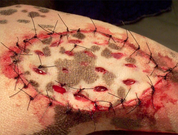
Meshed skin grafts have several advantages over non-meshed grafts.
Image courtesy Louise O’Dwyer.
Meshing is the creation of several rows of staggered, parallel incisions into a graft. Usually, a number 11 scalpel blade is used to make incisions 1cm long and roughly 1-2cm apart along the long axis of the graft.
The resultant mesh allows the graft to be stretched in two directions, increasing its flexibility and helping it conform to various shapes of wound.
For a given size of wound a smaller donor segment is required for meshed grafts because of the potential expansion, which may be helpful when a large wound requires reconstruction.
Small “pegs” of granulation tissue often grow into the meshed holes, providing further support and apposition between the graft and its bed. Also, meshed grafts allow passive drainage of fluid through the mesh holes, preventing accumulation of fluid under the graft, which could adversely affect graft take.
West Highland terrier being prepared for-x-ray. Image: Jackie Morrison
This view is particularly useful for demonstrating small pneumothoraces, emphysema or loculated pleural fluid.
The animal is placed in lateral recumbency. To demonstrate trapped pleural fluid, the affected side should be uppermost; to demonstrate emphysema, the affected side is placed down.
The x-ray cassette is positioned perpendicular to the spine on the table with a block – for example, a foam wedge or sandbag behind the cassette – to stop it from tipping backwards. The x-ray tube head is rotated so that it is parallel with the table and the x-ray beam is horizontal.
The x-ray beam should be directed towards an external wall and care should be taken that no people are on the other side of the wall.
One of the many non-academic challenges of becoming a vet is learning to cope with things not going to plan – to expect, or at least accept, the unexpected.
“We had planned to arrive in Agra to see the Taj Mahal on the one day a week it was closed…”
It may seem cliché to say travelling opens your eyes to different ways of life and changes you as a person, but the truth is it does prepare you for when the s*** hits the fan.
My friend and I had arrived in India with some trepidation; both of us had had busy summers and so very little time to consider what lay ahead.
We spent two days seeing some sights and travelling to our final destination, which was a feat in itself. India is just absolute mayhem.
Going to Goa
We had already circled Mumbai with a taxi driver who had no idea where he was going, returned to the hostel in the nick of time to grab our luggage for an onwards flight, only to be dropped off at the wrong airport and realise we had got our flight time wrong (though, thankfully, in our favour), before settling into our apartment in Goa, ready to start our EMS placement.
Having struggled to get in touch with the hosting charity, it finally arranged for a driver to pick us up on our first day. Therefore, on arrival at the shelter the final thing we expected was to be sat down by the board members of the charity, questioned and told they had no placement for us; we were subject to some miscommunication, goodbye.
Change of plans
Startled, with panic rising, we were shipped off to another charity 30km away to see whether they could offer us an alternative (which, after hours of discussion, they couldn’t).
After two days of frantic emailing and frustrating phone calls (with both parties struggling to understand accents) we found a saviour and abruptly departed Goa to fly to Delhi to squeeze in a few more sights before starting over.
This was also not without its challenges – we ended up directing two rickshaw drivers using maps on a phone, as they had both agreed to take us but, in reality, had no idea where our destinations were.
We battled the infamous Indian sleeper trains and had planned to arrive in Agra to see the Taj Mahal on the one day a week it was closed (again, calling for a swift diversion of plans). We had also been scammed on flights and made a total hash of accommodation bookings, having had to make so many last minute changes.
EMS saviours
“We battled the infamous Indian sleeper trains…”
Almost at the end of our tethers, we finally arrived in Jaipur to start our quickly organised placement at the charity Help In Suffering, which had completely saved our skins in terms of finding a suitable EMS placement that would count towards our degrees.
When it seemed like nothing else could possibly go wrong, something inevitably did. Nevertheless, we pulled each other through and overcame several unexpected challenges, despite being very close to just getting on the next plane home.
Although we had some extra hurdles, I think travelling in India at all is a total minefield for anyone, but you just have to accept the disorder and embrace the madness.
It may have started out (and continued for a fair while) as a total nightmare, but I definitely think we will both be better prepared for mishaps and abrupt, last minute changes in future veterinary practice – after all, we have this to reminisce over and think “it could be worse – at least we’re not stranded in India.”
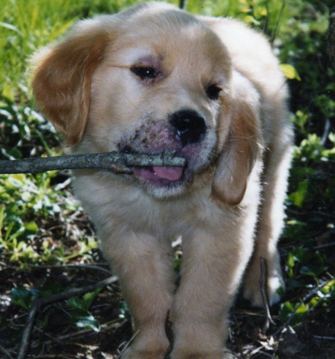
Golden retriever puppy with juvenile cellulitis. Photo: Trisha Shears, via Wikipedia.
Puppy pyoderma (also known as juvenile cellulitis) typically occurs between three weeks to four months of age, in any breed of puppy. It is characterised by an acute swelling of the face, especially the lips, eyelids, chin and muzzle.
It can be confused with an allergic reaction (e.g. a bee sting or vaccination reaction) and can develop into pustules that drain and scab.
The submandibular lymph nodes enlarge dramatically, hence giving the disease its other name of “strangles”.
Puppies are often lethargic, with appetite loss, pyrexia, and pain in the joints.
There may be an immune aetiology to the disease because affected animals respond well to steroids.
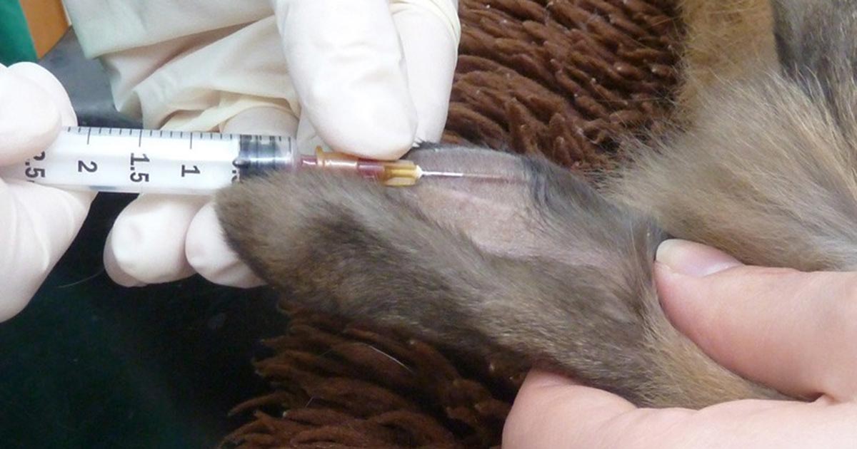
A venipuncture site should be chosen where the skin is clean and has no obvious inflammation or infection. The marginal ear vein or the lateral saphenous vein are usually good choices.
The fur should be clipped and the skin cleaned. EMLA cream can be applied over the site 45-60 minutes prior to venipuncture and covered with a dressing or cling film. The site should then be wiped with surgical spirit.
The vein is raised and the needle should be inserted at a very acute angle, almost parallel to the skin.
Gently does it…
Use only very gentle pressure to draw back on the syringe plunger, to prevent the vein from collapsing.
Too great a pressure may also damage the blood cells, especially when using a small gauge needle. Rotating the needle around its long axis so the bevel faces sideways or downwards may improve blood flow if the vein appears collapsed.
After taking the blood, pressure over the venipuncture site should be applied as rabbit veins are prone to haematomas.