Author: Kele Harris

  • Don’t do food trials in summer

    food-bowl-281980_640I now try to avoid running food trials in mid-summer. Certainly on first presentation, with no previous history of allergic dermatitis, I tend to treat accordingly and wait to see what happens later in the year as vegetation dies back.

    Food allergic dermatitis does not have a seasonal basis, so if the signs resolve or exacerbate over the course of the year, food allergy is not the primary cause (although some cases can confuse us as they have both an element of food allergy and atopic dermatitis).

    I have also seen cases started on food trials in the summer months that appear to get better as the year progresses, only for the owner to become reluctant to challenge as the dog is “better” – whereas, in reality, the improvement is the result of reduced exposure to an environmental allergen.

    So I usually wait and see if the signs persist to suggest a non-seasonal allergic dermatitis, and THEN do a food trial.


  • Immunotherapy in atopic dogs is a valuable tool

    Canine atopy
    “Canine Atopy” with dermatitis around the eye, caused by rubbing. Via Wikipedia Commons.

    My colleagues and I use immunotherapy on a fair number of dogs to treat atopic dermatitis. Research tells us it can be a valuable tool in some dogs, but at the same time up to a third of dogs respond poorly.

    We are lucky in our practice as anecdotally we find the majority of dogs do well, and we only get 10% to 15% that do not respond. This may be because:

    • We are really hot on parasite control in our practice – we have a large urban fox population and see cases of angiostrongylosis – so we advise Advocate on a regular, monthly basis. So it is unusual for us to see atopic dogs that flare due to concurrent parasite infestation.
    • We strongly advise owners to do and/or preserve with food trials – and some, okay, just one or two, do respond and relapse on challenge.
    • We also try to make sure we keep the perpetuating factors, for example, pyoderma and Malassezia dermatitis, to a minimum with medicated wipes and shampoos.
    • The older the dog when it starts immunotherapy the less likely it is to respond and we advise our owners accordingly.

    Good luck with those frustrating allergy cases – hopefully there are some tips here to help. And cats also do well on immunotherapy.

  • Using cytology to check for Malassezia organisms

    Using cytology to check for Malassezia organisms

    Fed up with your tape going all soggy?
    Fed up with your tape going all soggy?

    If, like me, you get frustrated using scotch tape in the Diff-Quik to check for Malassezia, I recently came up with a tip to avoid the tape going all soggy and cloudy in the fixative solution prior to staining.

    Take the strip as normal, but stick it directly onto a microscope slide. Then, using a 1ml syringe, collect a small amount of the final purple stain and inject it directly under the tape. Leave for a few minutes and then examine under oil immersion as normal.

    In my experience, this works just as well as the usual process.

  • “Hot water, soap and a towel please”

    Image ©iStockphoto.com/milosluz
    Image ©iStockphoto.com/milosluz

    This line always makes me think of the James Herriot books – and I recently thought of this when I had a poor cavalier King Charles that presented with a colon impacted with numerous shards of cooked lamb bones.

    The poor chap was in quite a bit of discomfort and was straining without any production. It took ages to flush out his colon and, to be honest, more than one attempt.

    Eventually some warm soapy water and repeated flushing with a cut dog-urinary catheter did the trick and he’s now feeling much better.

  • Use spay hooks to reveal pharyngeal foreign bodies in cats

    Use spay hooks to reveal pharyngeal foreign bodies in cats

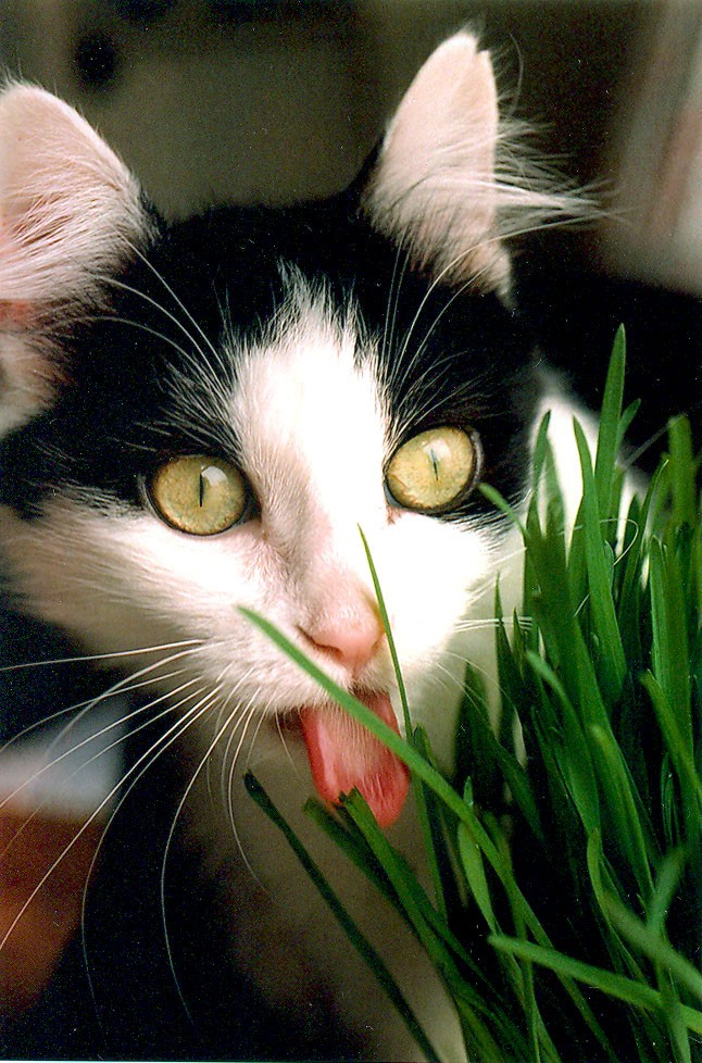
    Cat eating grass
    Image by Nancy Wong (Own work) [CC-BY-SA-3.0], via Wikimedia Commons.
    I find retching cats usually present last thing in an evening surgery, leaving you with that tricky choice of whether to anaesthetise and investigate immediately, or delay until the next morning.

    It also seems to be the ones you delay (because they’ve eaten, of course) that do have a grass blade while, more often than not, the ones you investigate there and then don’t.

    Anything there?

    A young colleague showed me a nifty way of elevating the soft palate with a spay hook, which neatly reveals the presence of grass (or not, as the case may be).

    Some careful work with the crocodile forceps and you can usually whip out the offending blade of grass very easily.

    Definitely a technique I’d recommend.

  • UPC – a good prognostic tool

    600px-Urine_sample
    Image courtesy Turbotorque

    Do you ever get blind spots, where you forget a particular test that might be useful as a prognostic indicator?

    I used to forget about the usefulness of urinary protein/creatinine ratios… until recently that is.

    To be honest, I think it may have been the need to collect a sample, package it up and send it off that sent this test scurrying into my subconscious.

    However, we now are able to run this in house and, as a result, we are running the test much more frequently – among all vets.

    This has really helped approach those renal cases in a rational way, both to give the owners an idea of prognosis and to decide when to institute a ACE inhibitor too.

  • Sarcoptes ELISA

    Sarcoptes scabei
    Sarcoptes scabei – a parasitic arthropod that burrows into skin and causes scabies. Image by Kalumet. Licensed under CC BY-SA 3.0 via Wikimedia Commons.

    Have you ever had an atopic dog that’s really well managed but suddenly flares, becomes very pruritic and seems to stop responding to therapy? I had one such case just a couple of months ago.

    Skin scrapes did not reveal the presence of any ectoparasites and cytology was pretty unexciting (a bit of bacterial colonisation but otherwise unremarkable).

    However, noticing the pruritus to be primarily around the head, ears and ventral abdomen tipped me off to think about Sarcoptes scabei – particularly as we have a very prolific and confident urban fox population in our practice area.

    A serum sample was duly sent off and – lo and behold – proved positive for Sarcoptes antibodies.

    The client then confessed to missing “a dose” of Advocate but, upon reapplication, management was readily restored.

  • It’s worth ruling out hypothyroidism if blood cholesterol is elevated

    Characteristic changes in the facial skin of a Labrador retriever with hypothyroidism.
    Characteristic changes in the facial skin of a Labrador retriever with hypothyroidism. Image by Caroldermoid (own work) [CC-BY-SA-3.0], via Wikimedia Commons.
    To be truthful, I have a bit of a blind spot when it comes to hypothyroidism. I often find few dogs follow the traditional textbook description of the clinical signs.

    I have seen two cases of late with elevated cholesterol, but no hair loss, unexplained weight gain, heat seeking and mucinous skin thickness.

    Both cases had blood submitted for total thyroxine (TT4) and thyroid-stimulating hormone (TSH), and both had very low TT4 levels and markedly increased TSH.

    Both cases are also improving nicely on thyroxine supplementation.

  • Neuro-exams can be very helpful

    Prolapsed gland of the third eyelid, or Cherry eye as it is sometimes known. Image by Joel Mills (CC BY-SA 3.0) via Wikipedia.
    Prolapsed gland of the third eyelid, or cherry eye as it is sometimes known. Image by Joel Mills (CC BY-SA 3.0) via Wikipedia.

    A nice six-year-old Labrador with a history of possible trauma, complete forelimb muscle atrophy and proprioceptive deficits was brought to see me.

    Radial paralysis was high up on the list of differentials, but the poor chap had not shown any response to conservative management.

    I noticed it had mild anisocoria, slight ptosis and third eyelid prolapse, indicating the less obvious Horner’s syndrome. A bell rang in my mind of a case I recalled seeing as a student – it revealed itself to be indicative of a brachial plexus injury/tumour.

    Sadly, an MRI scan at Davies Veterinary Specialists confirmed the presence of a tumour.

    Very sad.

  • Very blocked feline bladder

    I had a poor feline with a very blocked proximal urethra, and numerous attempts to pass various kinds of [feline] urinary catheter proved unfruitful.

    I’m indebted to a young colleague who helped by using two stay sutures through the prepuce to hold the penis in position – this puss was in good body condition, and this really helped expose and stabilise the penis.

    My colleague then used a 22G venous catheter inserted to the level of urolith, and a bit of hydro-propulsion, and we had easily passed a catheter.

    I must remember that one – thanks Dan (Preece).

    Image and description courtesy Danielle Gunn-Moore, BSc BVM&S PhD FHEA MANZCVSc, MRCVS, RCVS Specialist in Feline Medicine.
    Image & catheter options courtesy Danielle Gunn-Moore, BSc BVM&S PhD FHEA MANZCVSc, MRCVS, RCVS Specialist in Feline Medicine.


    Catheter options:
    The standard (Jackson-type) tomcat catheter (A) is stiff with a metal stylet and has rough side holes, so is traumatic. 

    The Slippery Sam (B) has end hole or side holes, is atraumatic and stiff, so good for initial unblocking. The Little Herbert adapter (C) is also shown as it makes it easier when attaching the catheter (B) to a closed collection system. 

    The hub of the Slippery Sam catheter is not firmly attached to the insertion tube, so it is recommended not to be left in situ more than six hours. The Fioniavet tomcat catheter (D) has an end hole, is atraumatic and stiff, so good for initial unblocking.